
CT and MRI Courses
Recent Course Feedback
Really informative and interesting course! Looking forward to part 2
Caroline G. Fundamental Skills in CT Course
All aspects explained really well. Applications and parameters of the scanner now make sense
Chloe V. Fundamental Skills in CT Course
Liz and Edward were very approachable. Felt no pressure. Explained with examples and drawings. Included us in group discussion. Great session!
Sarmita R. Fundamental Skills in MRI Course
I have worked in MRI for about 1.5 years now and this course has been very beneficial so far. It has helped to understand the equipment better and to be less afraid of playing with parameters within reasonable limits
Madara D. Fundamental Skills in MRI Course
I just want to say thankyou to Liz and Edward for this fantastic course. You were very thorough and covered the majority of the areas a newbie into MRI like myself found very overwhelming and daunting when I initially tried studying the physics aspect on my own. So once again, thank you both very much
Anita K. Fundamental Skills in MRI Course

USEFUL LINKS
MSK CASES
www.mskradiology4u.co.uk
MSK Radiology Signs
http://www.gentili.net/signs
See our current course listings on:
The CSP website: www.csp.org.uk/events

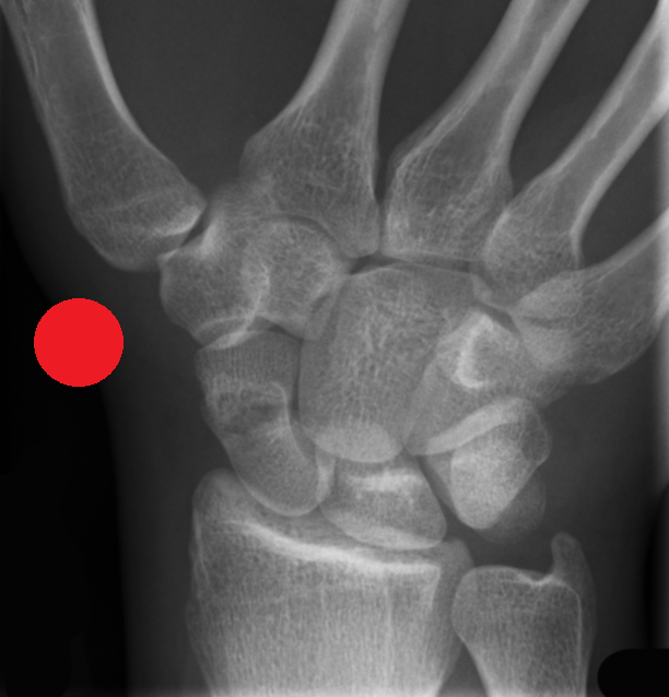
Red Dot X-Ray Interpretation Course (Online)

Course is College of Radiographers CPD Now Endorsed
Course Focus
This Red Dot X-Ray Interpretation Course is designed for Advanced Clinical Practitioners and Radiographers working in acute MSK care, for example in A/E or minor injuries and who are seeking to enhance confidence and improve their diagnostic assessment and accuracy. Advanced Clinical Practitioners eligible for attendance include Advanced Nurse Practitioners and Advanced Physiotherapist Practitioners.
The course focuses on the identification and escalation of suspected pathology, fractures, or other deviations from normal imaging appearances. Participants will build robust knowledge and improve their ability to flag potentially critical findings.
The course will support advanced critical thinking to facilitate delivery of safe and competent care for patients. It is suitable for individuals who are currently working as an ACP or Radiographer together with those working towards these roles. The course will also support and complement existing education required to practice as an Advanced Clinical Practitioner eg. ANP/APP.
What is COR CPD Now Endorsement?
The course has been reviewed by the COR's Approval and Accreditation Board. A CPD Now Endorsed course is a confirmation that the training aligns with professional, evidenced based practice and meets HCPC (Health Care Professions Council) Standards, providing a quality mark and confidence that you are attending a course of the highest quality.
The course has been measured against the following Learning Outcomes:
CoR 01: Practical Skills
CoR 02: Knowledge Base
CoR 03: Work Safely
CoR 04: Legal/ethical frameworks or guidance
CoR 05: Communication Skills
CoR 19: Evidence to Support Practice
Course Content and Learning Outcomes
The course focuses on improving the skills needed to identify a range of acute pathologies on x-rays, including fractures, dislocations, chest abnormalities and more. The course will improve patient safety by reducing missed urgent findings, especially whilst waiting for report completion.
On successful completion, delegates will exhibit enhanced practice in the following areas:
- Explanation of the principles and purpose of the red dot system in radiology, it's integration into clinical pathways and it's role in the early identification of acute abnormalities.
- Appreciate the impact of early abnormality detection on patient outcomes, workflow efficiency and overall quality of care.
- Improved identification of key radiographic features of common acute pathologies, eg. fractures, dislocations, pnemothorax.
- Improved systematic image review techniques and use of structured approaches to review radiographic images and minimise diagnostic oversight.
- Improved ability to distinguish between normal anatomical variants and common pathological findings on Radiographs.
- Demonstrate clear and timely communication of urgent findings to the appropriate clinical teams, adhering to best practices in documentation and escalation.
- Recognise the limitations of the red dot system and understand when to seek further radiological or clinical input.
- Awareness of professional roles in the red dot system and the governance framework.
- Critically evaluate personal performance in image interpretation.
Requirements for Course Access and Completion
Course access will require a PC/Mac desktop or laptop - minimum 14 inch screen, suitable for viewing and assessing images. Delegates will need to access the course from a quiet, lone working location without interruption due to the assessment on day two. A working webcam and microphone is also essential. Delegates will be unable to undertake the assessment if the above requirements are unable to be met.
Course Suitability
Suitable for Current Advanced Clinical Practitioners including Advanced Nurse Practitioners and Advanced Physiotherapist Practitioners together with Radiographers. In order to complete the logbook component, the delegate will need to have access to urgent/trauma cases and be currently rotating through an urgent care setup, for example A/E or a minor injuries unit.
The course is also open to delegates working towards these roles. Please contact us for further details.
Course Delivery
The course is delivered online, is live and interactive and is designed to be attended by delegates from multiple disciplines involved in the care and management of patients in acute MSK care. An online image viewing assessment is included together with a requirement to complete a logbook of cases and a post course reflective piece of work.
The course is split into:
a) Theoretical Study (Ongoing evidenced theoretical knowledge - continual micro assessments).
b) Logbook (independent reflective practice in the delegate's department).
c) Assessment (evidenced application of theoretical knowledge and evidenced pattern recognition skills).
d) Reflective Writing (awareness of application and impact).
More information on the course leads can be found here:
Course Programme
Day 1 (six hours contact time)
- Theory of Pattern Recognition and the Red Dot System.
- Application in the Axial Skeleton.
- Application in the Appendicular Skeleton.
Between Day 1 and Day 2: Completion of a 15 case logbook in delegate's department (15 cases). This will include:
- Basic clinical history of the case.
- Whether or not a red dot was applied to the case.
- Review of decision by peer/senior colleague, as appropriate.
- Whether peer/senior colleague agreed or disagreed with delegate's decision.
- Perceived difficulty level 1-5 (by delegate) of the case.
- Comments by senior colleague and delegate, identifying any areas for improvement.
Day 2 (six hours contact time)
- Logbook Reviews.
- Image Theory Recap - Quiz.
- Red Flag and Incidental Findings.
- Image Viewing Preparation for Assessment (Variety of Axial and Appendicular Skeleton).
- Image Viewing Assessment (10 Axial, 10 Appendicular Cases).

Book Your Place

Spring Course 2026
8th May (Part 1)
22nd May (Part 2)
£325+VAT
Zoom Waiting Room from 08:50am.
Registration and Welcome 09:00am.
Course Times:
09:00-16:00 Part 1
09:00-16:00 Part 2
Minimum 12 Hours Contact Time